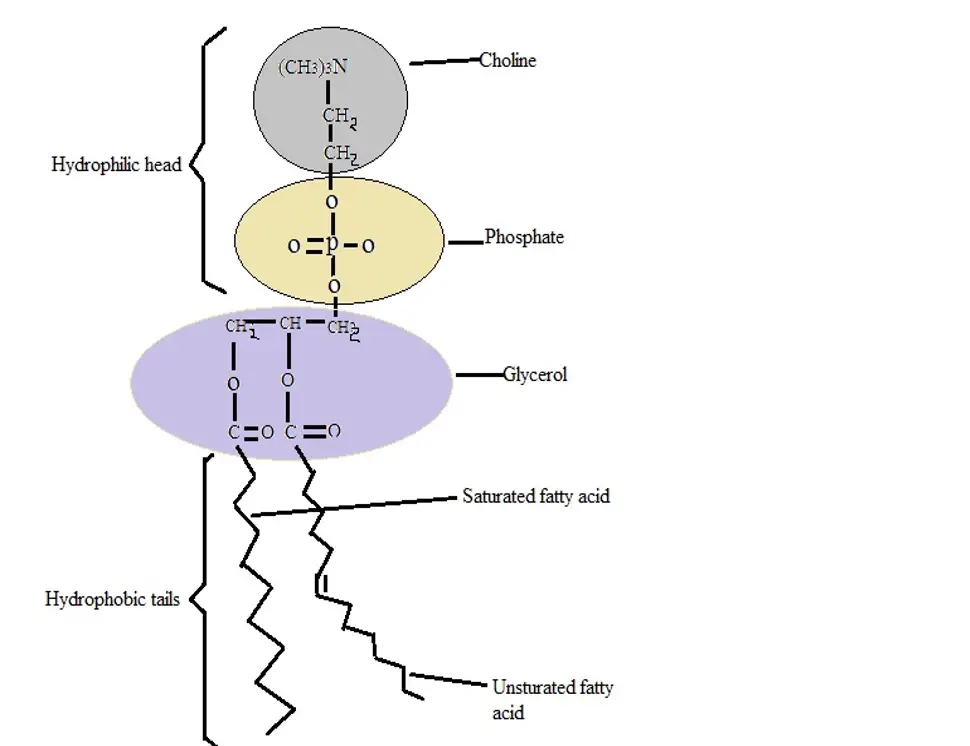
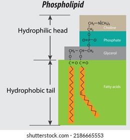
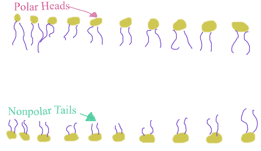
45 drawing of a phospholipid
Notes CELL STRUCTURE AND FUNCTION - National Institute of … z illustrate the structure of plant and animal cells by drawing labelled diagrams; z describe the structure and functions of plasma membrane, cell wall, endoplasmic reticulum (ER), cilia, flagella, nucleus, ribosomes, mitochondria, chloroplasts, golgi body, peroxisome, glyoxysome and lysosome; z describe the general importance of the cell molecules-water, mineral ions, … 117054: Lupus Anticoagulant Comprehensive | Labcorp Lupus anticoagulants are antibodies that inhibit one or more of the in vitro phospholipid-dependent tests of coagulation. 6-10 Recently, the SCC Subcommittee for the Standardization of Lupus Anticoagulants provided guidelines for the laboratory diagnosis of LA. 6 No single screening test can detect all LA-positive patients. The ISTH recommends that any sample …
Cells - University of Utah In multicellular organisms, cells work together in teams. Multiple cell types, each specialized for a certain function, team up to form tissues.

Drawing of a phospholipid
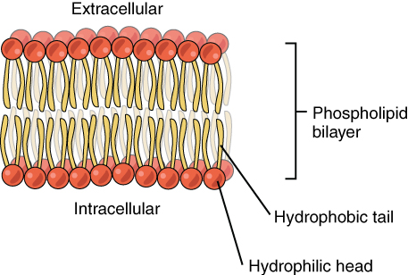
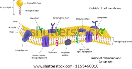
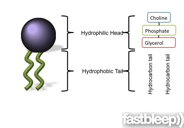
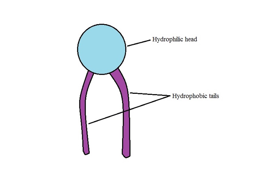
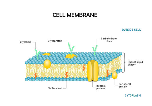
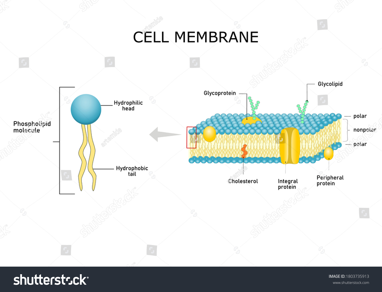
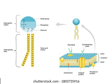
2.3 Cell structure and function | Cells: the basic units of ... Each phospholipid has a polar, hydrophilic (water-soluble) head as well as a non-polar, hydrophobic (water-insoluble) tail. It is a semi-permeable structure that does not allow materials to pass through the membrane freely, thus protecting the intra and extracellular environments of the cell. Organic & Biomolecular Chemistry Ruthenium photoredox-triggered phospholipid membrane formation From DOI: 10.1039/C6OB00290K. An alternative, unsuitable way of writing the final example above would be: Ru(bpy 3) 2+ 11-watt CFL-triggered tool reduces Cu(II) and initiates CuAAC reaction leading to phospholipid synthesis and in situ membrane formation. This title is too detailed and includes … Blood Specimens: Coagulation | Labcorp Laxson CJ, Titler MG. Drawing coagulation studies from arterial lines; an integrative literature review. Am J Critical Care. 1994 Jan; 3(1):16-24. Lew JK, Hutchinson R, Lin ES. Intra-arterial blood sampling for clotting studies. Effects of heparin contamination. Anaesthesia. 1991 Sep; 46(9):719-721.
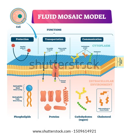
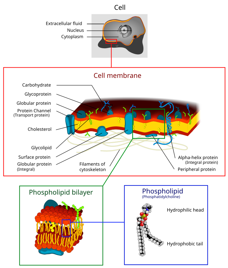
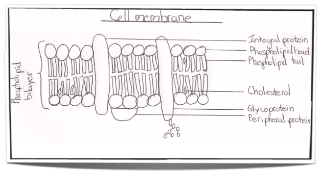
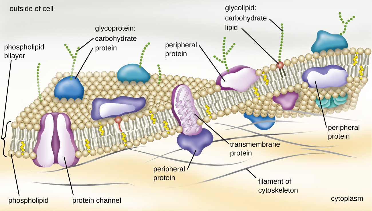
Drawing of a phospholipid. AQA A Level Biology 4/5/6 mark questions Flashcards | Quizlet Double layer of phospholipid molecules; 2. Detail of arrangement of phospholipids; 3. Intrinsic proteins/protein molecules passing right through; 4. Some with channels/pores; 5. Extrinsic proteins/proteins only in one layer/on surface; 6. Molecules can move in membrane/dynamic/membrane contains cholesterol; 7. Glycocalyx/carbohydrates attached to … Describe the biological need for cells to be surrounded by a … 10 2-fold difference for other ions such as Na +, K +, and Mg 2+) (30, 31). It is often the largest organelle in the cell. Cytosol is the fluid of Cytoplasm Cell Membrane and Transport: Learn how transporters keep cells healthy Virtual Lab. Membranes not only separate different areas but also control All cells are surrounded by a cell membrane that ... Lipids - Michigan State University If a phospholipid is smeared over a small hole in a thin piece of plastic immersed in water, a stable planar bilayer of phospholipid molecules is created at the hole. As shown in the following diagram, the polar head groups on the faces of the bilayer contact water, and the hydrophobic alkyl chains form a nonpolar interior. The phospholipid molecules can move about in their half … Cells - University of Utah In multicellular organisms, cells work together in teams. Multiple cell types, each specialized for a certain function, team up to form tissues.
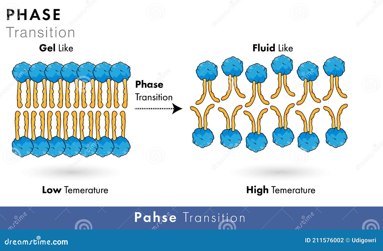
Lipids - Michigan State University The phospholipid molecules can move about in their half the bilayer, but there is a significant energy barrier preventing migration to the other side of the bilayer. To see an enlarged segment of a phospholipid bilayer Click Here. This bilayer membrane structure is also found in aggregate structures called liposomes. Liposomes are microscopic ... AQA A Level Biology 4/5/6 mark questions Flashcards | Quizlet 1. Double layer of phospholipid molecules; 2. Detail of arrangement of phospholipids; 3. Intrinsic proteins/protein molecules passing right through; 4. Some with channels/pores; 5. Extrinsic proteins/proteins only in one layer/on surface; 6. Molecules can move in membrane/dynamic/membrane contains cholesterol; 7. 2.3 Cell structure and function | Cells: the basic units of life - Siyavula The lipid bilayer forms spontaneously due to the properties of the phospholipid molecules. In an aqueous environment, the polar heads try to form hydrogen bonds with the water, while the non-polar tails try to escape from the water. The problem is solved by the formation of a bilayer because the hydrophilic heads can point outwards and from hydrogen bonds with water, and … Prokaryotic and Eukaryotic Cells Flashcards | Quizlet 4) The best definition of osmotic pressure is A) The movement of solute molecules from a higher to a lower concentration. B) The force with which a solvent moves across a semipermeable membrane from a higher to a lower concentration.
Blood Specimens: Coagulation | Labcorp Lupus anticoagulants (LA) are are nonspecific antibodies that extend clot-based coagulation assays as the result of their interaction with phospholipid in the reaction mixture. Platelets in plasma samples can act as a source of phospholipid and mask the effects of LA. For this reason, it is important to prepare platelet-poor plasma (PPP) for LA testing. PPP should have a platelet … 117054: Lupus Anticoagulant Comprehensive | Labcorp Lupus anticoagulants are antibodies that inhibit one or more of the in vitro phospholipid-dependent tests of coagulation. 6-10 Recently, the SCC Subcommittee for the Standardization of Lupus Anticoagulants provided guidelines for the laboratory diagnosis of LA. 6 No single screening test can detect all LA-positive patients. Prokaryotic and Eukaryotic Cells Flashcards | Quizlet Start studying Prokaryotic and Eukaryotic Cells. Learn vocabulary, terms, and more with flashcards, games, and other study tools. Blood Specimens: Coagulation | Labcorp Laxson CJ, Titler MG. Drawing coagulation studies from arterial lines; an integrative literature review. Am J Critical Care. 1994 Jan; 3(1):16-24. Lew JK, Hutchinson R, Lin ES. Intra-arterial blood sampling for clotting studies. Effects of heparin contamination. Anaesthesia. 1991 Sep; 46(9):719-721.
Organic & Biomolecular Chemistry Ruthenium photoredox-triggered phospholipid membrane formation From DOI: 10.1039/C6OB00290K. An alternative, unsuitable way of writing the final example above would be: Ru(bpy 3) 2+ 11-watt CFL-triggered tool reduces Cu(II) and initiates CuAAC reaction leading to phospholipid synthesis and in situ membrane formation. This title is too detailed and includes …
2.3 Cell structure and function | Cells: the basic units of ... Each phospholipid has a polar, hydrophilic (water-soluble) head as well as a non-polar, hydrophobic (water-insoluble) tail. It is a semi-permeable structure that does not allow materials to pass through the membrane freely, thus protecting the intra and extracellular environments of the cell.













Post a Comment for "45 drawing of a phospholipid"