39 neuron diagram labeled
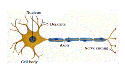
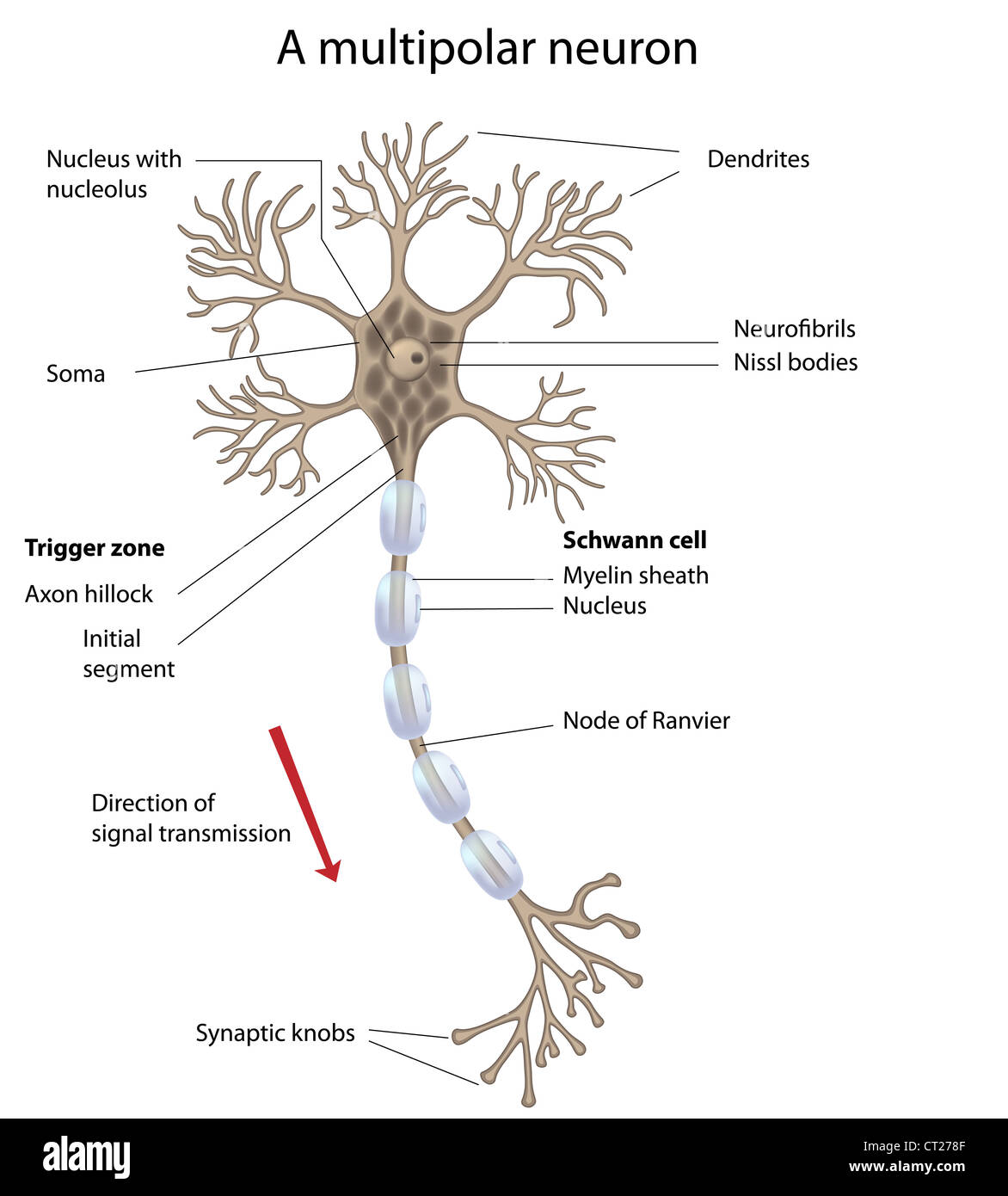
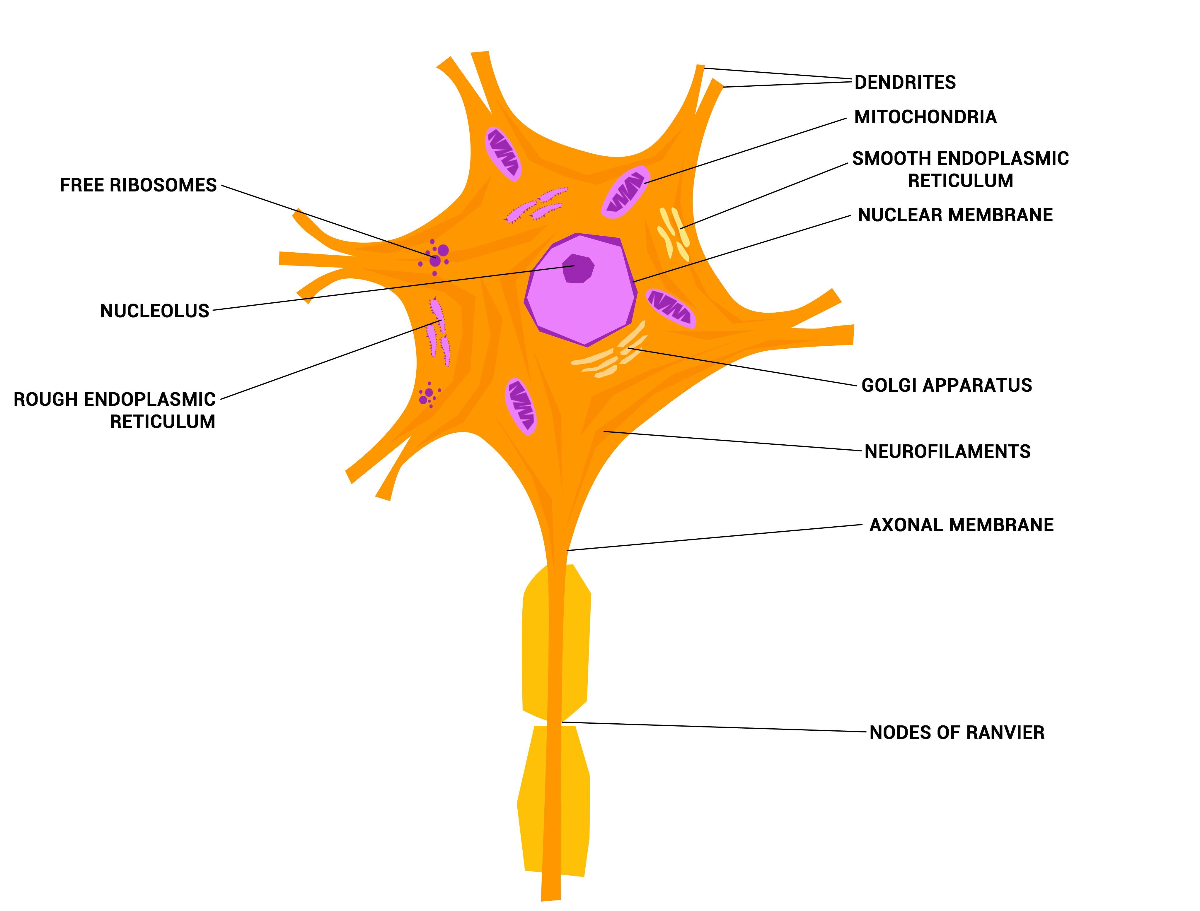
A Labelled Diagram of Neuron with Detailed decription - Collegedunia A neuron is a type of cell that is largely responsible for conveying information via electrical and chemical impulses. The brain, spinal cord, and peripheral nerves all contain them. The nerve cell is another name for a neuron. The structure of a neuron changes depending on its form and size, as well as its function and location. Describe the structure and function of neuron with labelled diagram ... Diagram of neuron with labels Here is the description of human neuron along with the diagram of the neuron and their parts. The neuron is a specialized and individual cell, which is also known as the nerve cell. A group of neurons forms a nerve.
how to draw structure of neuron/neuron diagram labelled ... - YouTube Please watch: "cell structure and functions / animal cell vs plant cell / parts of cell / ch 8 science class 8 cbse" ...

Neuron diagram labeled
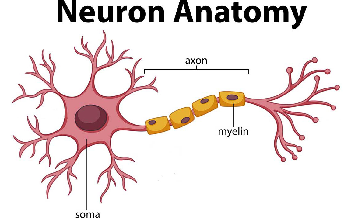
Label Neuron Anatomy Printout - EnchantedLearning.com Read the definitions, then label the neuron diagram below. axon - the long extension of a neuron that carries nerve impulses away from the body of the cell. axon terminals - the hair-like ends of the axon. cell body - the cell body of the neuron; it contains the nucleus (also called the soma) Labeled Neuron Diagram | Quizlet Start studying Labeled Neuron. Learn vocabulary, terms, and more with flashcards, games, and other study tools. Home. ... Essentials Of Human Anatomy Physiology And Modified Mastering A P With Pearson Etext -- Valuepack Access Card Package ( Edition) ... Brian Ventricles Diagram. 11 terms. Victoriaruanto PLUS. Sets with similar terms. Anatomy ... Neuron - Wikipedia A neuron or nerve cell is an electrically excitable cell that communicates with other cells via specialized connections called synapses. ... Diagram of a typical myelinated vertebrate motor neuron. ... Anatomy of a neuron; Neuron images This page was last edited on 15 September 2022, at 08:55 (UTC). Text is ...
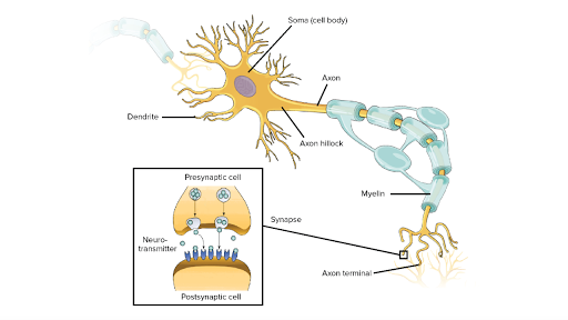
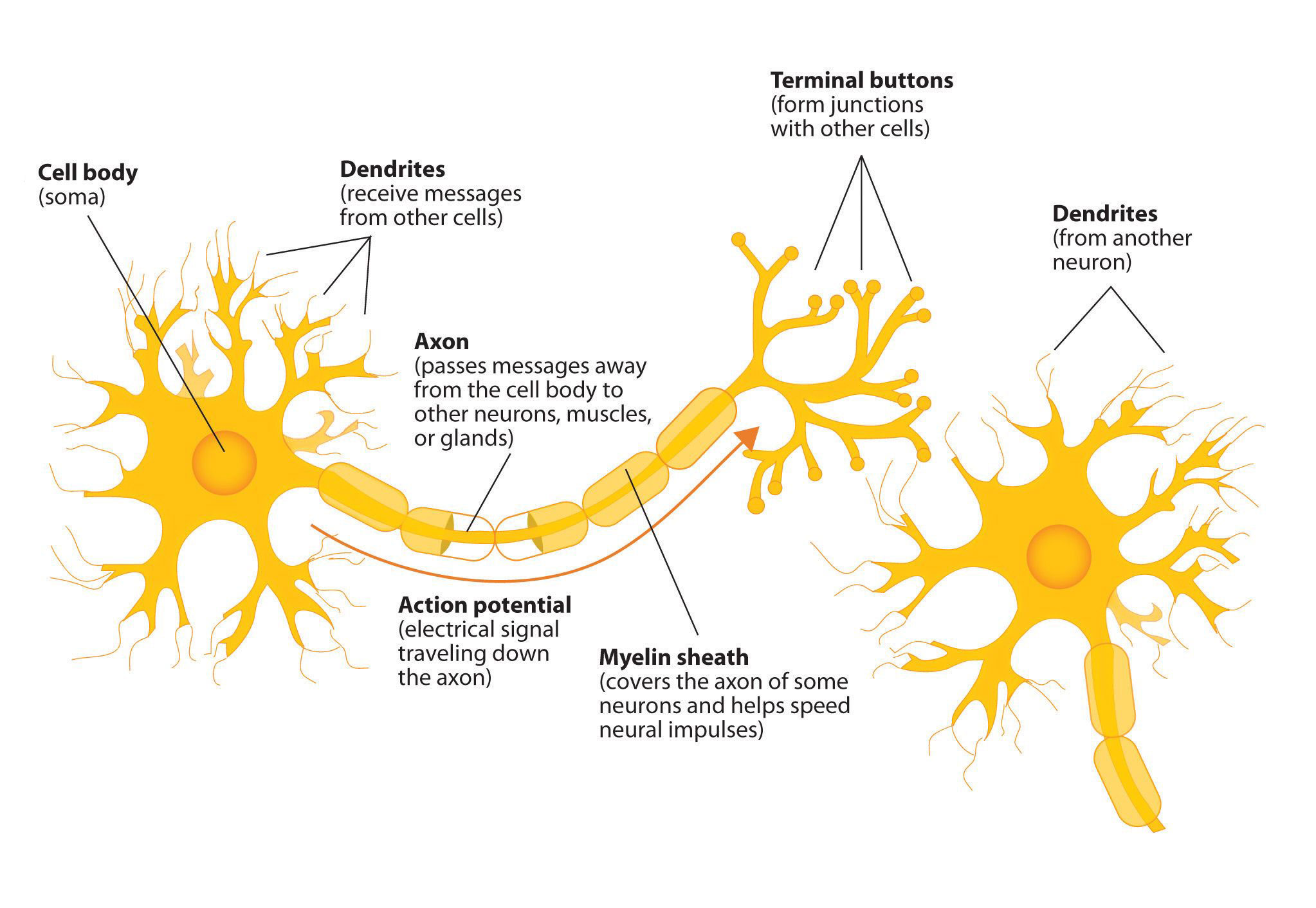
Neuron diagram labeled. Neuron Diagram & Types | Ask A Biologist - Arizona State University Neuron Anatomy. Nerve Cell: Dendrites receive messages from other neurons. The message then moves through the axon to the other end of the neuron, then to the tips of the axon and then into the space between neurons. From there the message can move to the next neuron. Neurons pass messages to each other using a special type of electrical signal. Nervous System - Label the Neuron - TheInspiredInstructor.com Nervous System - Neuron: Nerve Cell. Choose the correct names for the parts of the neuron. (6) This neuron part receives messages from other neurons. (7) This neuron part sends on messages to other neurons. (8) This neuron part gives messages to muscle tissue. (9) This neuron part processes incoming messages. Structure of Neurons: What Is a Neuron? Types, Structure, Parts Structure of Neuron. Each neuron has a cell body, which is the central area of the neuron. It contains the nucleus and other structures common to all cells in the body, such as mitochondria. Neurons have highly branched fibres that reach out from the neuron are called dendritic trees. Each branch is called a dendrite. Overview of neuron structure and function - Khan Academy The basic functions of a neuron. If you think about the roles of the three classes of neurons, you can make the generalization that all neurons have three basic functions. These are to: Receive signals (or information). Integrate incoming signals (to determine whether or not the information should be passed along).
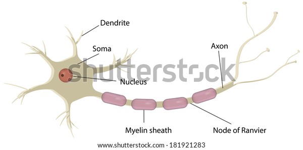
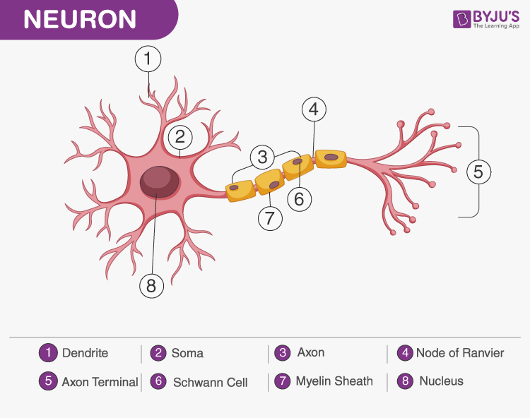
A Labelled Diagram Of Neuron with Detailed Explanations - BYJUS A Labelled Diagram Of Neuron with Detailed Explanations Biology Biology Article Diagram Of Neuron Diagram Of Neuron A neuron is a specialized cell, primarily involved in transmitting information through electrical and chemical signals. They are found in the brain, spinal cord and the peripheral nerves. A neuron is also known as the nerve cell. What Is a Neuron? Diagrams, Types, Function, and More - Healthline An Easy Guide to Neuron Anatomy with Diagrams Medically reviewed by Nancy Hammond, M.D. — By Carly Vandergriendt and Rachael Zimlich, RN, BSN — Updated on February 28, 2022 Neuron under Microscope with Labeled Diagram - AnatomyLearner Neuron under Microscope with Labeled Diagram 31/03/2022 31/03/2022 by anatomylearner The structural and functional unit of the nervous system is the neuron that may easily observe under a light microscope. Neurons may vary considerably in size, shape, and other features. File:Complete neuron cell diagram en.svg - Wikipedia English: Complete neuron cell diagram. Neurons (also known as neurones and nerve cells) are electrically excitable cells in the nervous system that process and transmit information. In vertebrate animals, neurons are the core components of the brain, spinal cord and peripheral nerves. Own work.
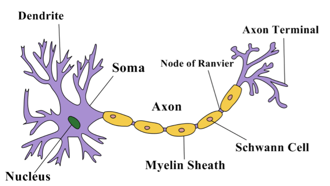
Labeled Neuron Diagram | Science Trends Neurons are a type of cell and are the fundamental constituents of the nervous system and brain. Neurons take in stimuli and convert them to electrical and chemical signals that are sent to our brain. There are 3 major kinds of neurons in the spinal cord: sensory, motor, and interneurons. Neurons communicate vie electrical signals produced by ... Neurons (With Diagram) - Biology Discussion A neuron is a structural and functional unit of the neural tissue and hence the neural system. Certain neurons may almost equal the length of body itself. Thus neurons with longer processes (projections) are the longest cells in the body. Human neural system has about 100 billion neurons. Majority of the neurons occur in the brain. Label Parts of a Neuron Diagram | Quizlet Dendrites. receives impulses from other nerve cells. axon hillock. The cell body...the part of the cell that houses the nucleus and keeps the entire cell alive and functioning. Myelin Sheath. Surrounds the axon an insulates it from surrounding cells and tissues and making signal transitions faster and more efficient. Terminal Buttons. Labeled Neuron Diagram in 2022 | Neuron diagram, Neurons, Science diagrams Labeled Neuron Diagram The following labeled diagram shows the parts of a neuron. In order to make it more understandable to the students, we have added all the functions of the Neuron in the labeled diagram. Wondershare Edraw 8k followers More information Labeled Neuron Diagram
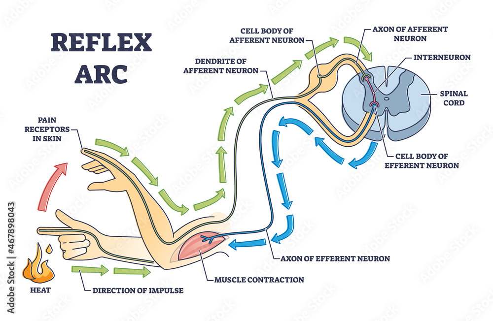
Labeled Diagram Of A Neuron Illustrations, Royalty-Free Vector ... - iStock Labeled diagram of the neuron Labeled diagram of the neuron, nerve cell that is the main part of the nervous system. labeled diagram of a neuron stock illustrations Labeled diagram of the neuron Reflex arc explanation with pain signals and receptor impulse outline diagram Reflex arc explanation with pain signals and receptor impulse outline diagram.
Diagram Quiz on Neuron Structure and Function (Labeling Quiz) This labelled diagram quiz on Neuron is designed to assess your basic knowledge in Structure and Function of Neuron. Choose the best answer from the four options given. When you've finished answering as many of the questions as you can, scroll down to the bottom of the page and check your answers by clicking ' Score '. Percentage score will be displayed along with right answers.
Neuron Diagram Teaching Resources | Teachers Pay Teachers Worksheets included: Neuron - Match names of neuron structures with labeled diagram Neuron 2 - Identify neuron structures on diagram and describe structure functions 2 answer keys are included. This product is also available as a part of a Nervous System Worksheet Pack with Diagrams We are actively adding content to our store. Please
Neuron Diagram Unlabeled Unlabeled diagram of a motor neuron (try labeling: axon, dendrite, cell body, myelin, nodes of Ranvier, motor end plate).Read the definitions, then label the neuron diagram below. axon - the long extension of a neuron that carries nerve impulses away from the body of the cell. axon terminals - the hair-like ends of the axon cell body - the cell ...
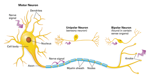
Types of Neurons: Parts, Structure, and Function - Verywell Health Summary. Neurons are responsible for transmitting signals throughout the body, a process that allows us to move and exist in the world around us. Different types of neurons include sensory, motor, and interneurons, as well as structurally-based neurons, which include unipolar, multipolar, bipolar, and pseudo-unipolar neurons.
Labeled Neuron Diagram - 24HTECH.ASIA Labeled Neuron Diagram. Neurons are the basic organizational units of the brain and nervous system. Neurons form the bulk of all nervous tissue and are what allow nervous tissue to conduct electrical signals that allow parts of the body to communicate with each other. Neurons are the cells that are responsible for receiving sensory input from ...
A Guide to Understand Neuron with Neuron Diagram 3. How to Draw a Neuron Diagram To learn about the structure of the neurons, the students can use a neuron labeled diagram. The students may follow these steps to make their neuron diagram, but the process is complex: 3.1 How to Draw a Neuron Diagram from Sketch Step 1: First, the students need to draw a circle. Based on it, they need to draw a star-like shape.
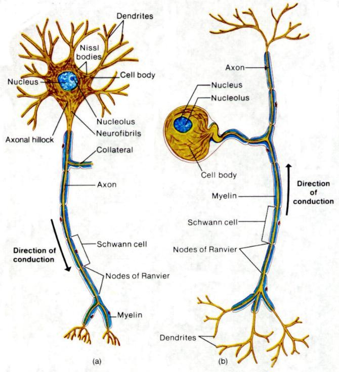
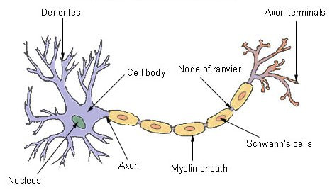
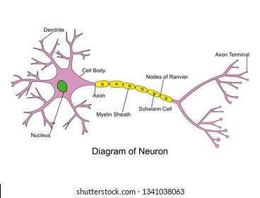
Neuron Diagram Labeled | EdrawMax Template It is an effective form of self-assessment, enabling students to check their understanding. In the following diagram, we have illustrated the important parts of the Neuron. In the following Neuron labeled diagram, we have dendrite, cell body, axon, myelin sheath, Schwann cell, a node of Ranvier, axon terminal, and nucleus.
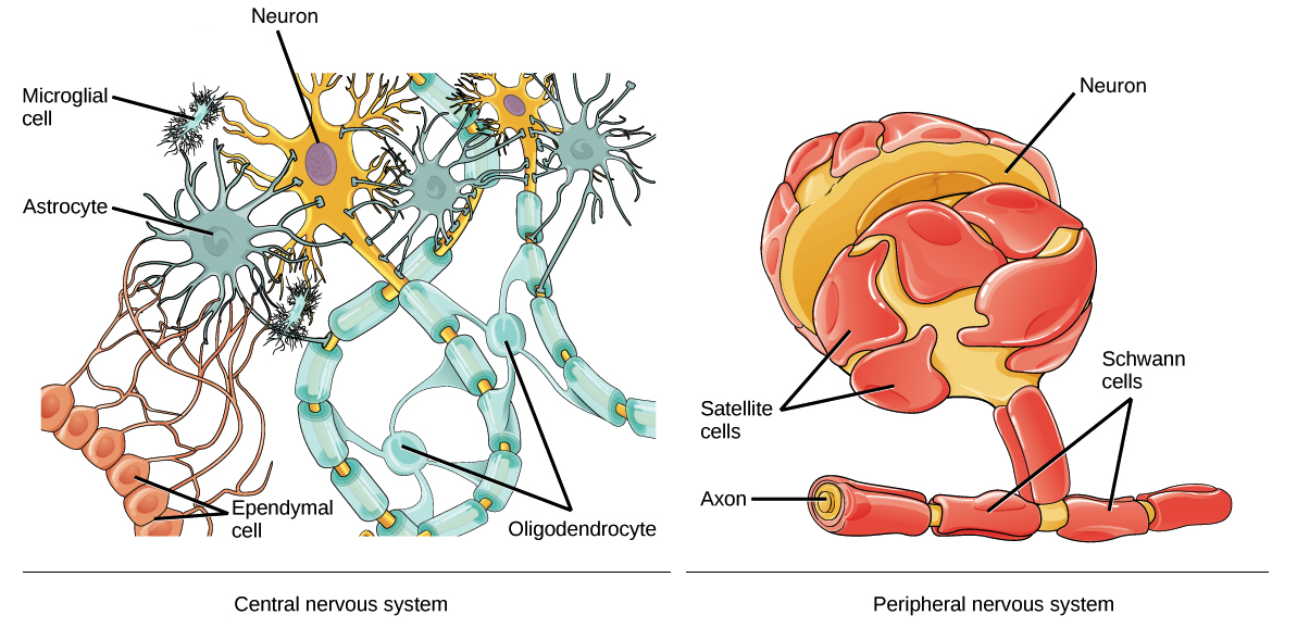
Sensory Neuron Diagram Illustrations & Vectors - Dreamstime Labeled diagram of the Neuron, nerve cell that is the main part of the nervous system. Abstract grey mesh background. Cells of human`s brain. Neuron and glial cells Microglia, astro. Cyte and oligodendrocyte. Vector diagram for educational, medical, biological and science use.
Location, Structure, and Functions of Motor Neurons - Bodytomy Being the most basic units of the human nervous system, neurons play a vital role in sensing and responding to different external as well as internal stimuli. A motor neuron is one of the three types of neurons involved in this process. Read about the structure and function of a motor neuron with reference to a neatly labeled diagram, in this Bodytomy post.
An Easy Guide to Neuron Anatomy with Diagrams - SimplyPsychology.org Neurons are the information processing units of the brain which have a responsibility for sending, receiving, and transmitting electrochemical signals throughout the body. Neurons, also known as nerve cells, are essentially the cells that make up the brain and the nervous system. Neurons do not touch each other, but where one neuron comes close ...
Neuron - Wikipedia A neuron or nerve cell is an electrically excitable cell that communicates with other cells via specialized connections called synapses. ... Diagram of a typical myelinated vertebrate motor neuron. ... Anatomy of a neuron; Neuron images This page was last edited on 15 September 2022, at 08:55 (UTC). Text is ...
Labeled Neuron Diagram | Quizlet Start studying Labeled Neuron. Learn vocabulary, terms, and more with flashcards, games, and other study tools. Home. ... Essentials Of Human Anatomy Physiology And Modified Mastering A P With Pearson Etext -- Valuepack Access Card Package ( Edition) ... Brian Ventricles Diagram. 11 terms. Victoriaruanto PLUS. Sets with similar terms. Anatomy ...
Label Neuron Anatomy Printout - EnchantedLearning.com Read the definitions, then label the neuron diagram below. axon - the long extension of a neuron that carries nerve impulses away from the body of the cell. axon terminals - the hair-like ends of the axon. cell body - the cell body of the neuron; it contains the nucleus (also called the soma)












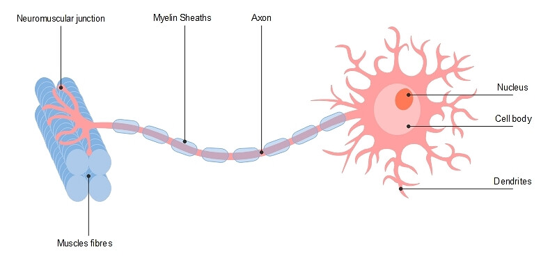
Post a Comment for "39 neuron diagram labeled"