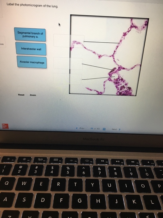
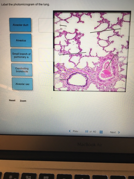
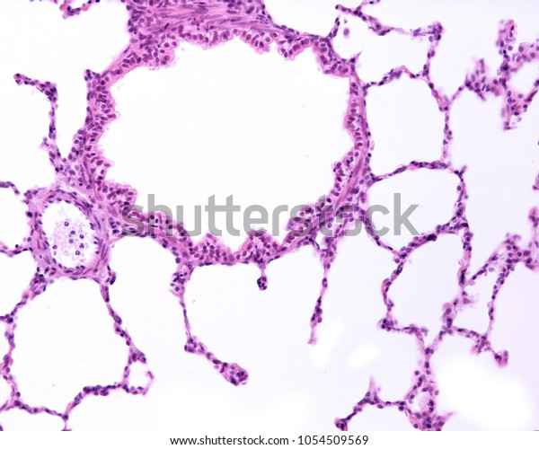
43 label the photomicrogram of the lung
The Lungs - Position - Structure - TeachMeAnatomy There are three lung surfaces, each corresponding to an area of the thorax. The mediastinal surface of the lung faces the lateral aspect of the middle mediastinum. The lung hilum (where structures enter and leave the lung) is located on this surface. The base of the lung is formed by the diaphragmatic surface. It rests on the dome of the diaphragm, and has a concave shape. EOF
Labeled diagram of the lungs/respiratory system. - SERC Labeled diagram of the lungs/respiratory system. Image 37789 is a 1125 by 1408 pixel PNG. Uploaded: Jan10 14. Last Modified: 2014-01-10 12:15:34. Permanent URL: . The file is referred to in 1 page. Airborne Microbes.

Label the photomicrogram of the lung
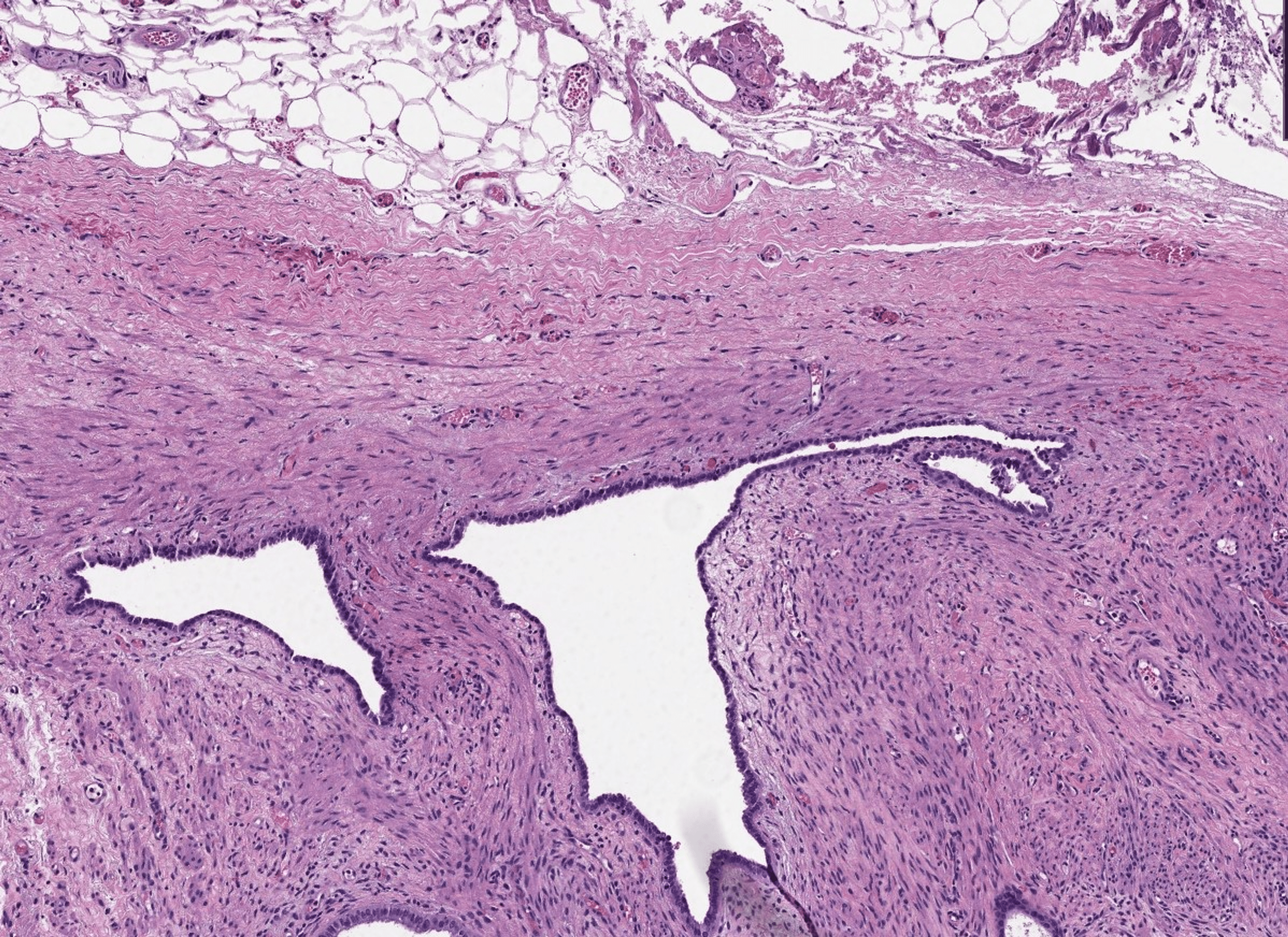
Label the photomicrogram of the trachea. - Brainly.com Label the photomicrogram of the trachea. Get the answers you need, now! mooretami1829 mooretami1829 07/20/2022 SAT High School answered • expert verified Label the photomicrogram of the trachea. 1 See answer Advertisement Photomicrograph of the affected lung lobe, showing the severe ... Download scientific diagram | Photomicrograph of the affected lung lobe, showing the severe emphysema. Smooth muscle hyperplasia is present in some alveolar walls, and around the bronchiole (upper ... A&P 2 Lab Unit 2 Flashcards | Quizlet Label the photomicrogram of the lung. Identify the cartilaginous anatomical structures shown in the posterior view of the superior portion of the lower respiratory system. Place the following structures with the appropriate anatomical region. Labels maybe be placed in more than one category.
Label the photomicrogram of the lung. A&P 2 lab ex 33 Flashcards | Quizlet Start studying A&P 2 lab ex 33. Learn vocabulary, terms, and more with flashcards, games, and other study tools. Solved Label the photomicrogram of the lung Alveolar duct - Chegg A.Alveolar duct. B.SMALL BRANCH OF PULMONARY A …. View the full answer. Transcribed image text: Label the photomicrogram of the lung Alveolar duct Alveolus Small branch of pulmonary a. bronchiole Alveolar sac Reset Zoom < Prev 22 of 40::: Next > 20. A&P 139 Chapter 19 Flashcards | Quizlet Study with Quizlet and memorize flashcards terms like common passageway for air and food; passageway for air only, A decrease in surface area and decrease in gas exchange., false and more. Lung Anatomy, Function, and Diagrams - Healthline The trachea and bronchi airways form an upside-down "Y" in your chest. This "Y" is often called the bronchial tree. The bronchi branch off into smaller bronchi and even smaller tubes ...
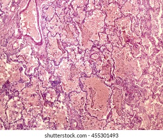
Photomicrograph of the Lung Quiz - PurposeGames.com Games by same creator. 2p Image Quiz. 8p Image Quiz. Reproductive Systems 2 games. The Integumentary System. The Nervous System & Senses. The Digestive System. The Human Body-- An Orientation 7 games. The Microscope 2 games. Label The Photomicrograph Of The Lung : 4 Chloro Dl Phenylalanine ... Label the photomicrogram of the lung segmental branch of pulmonary a. Make sure you know the basics of lung cancer, including prevention, risk factors, symptoms and treatment options. Label the anterior view of the lower respiratory tract based on the hints if. Electron micrograph of lung tissue (click to show / hide labels). Photomicrograph of the Lung Quiz - purposegames.com This online quiz is called Photomicrograph of the Lung. This game is part of a tournament. You need to be a group member to play the tournament Label the lung diagram Diagram | Quizlet Start studying Label the lung diagram. Learn vocabulary, terms, and more with flashcards, games, and other study tools. Svg Vector Icons :
Answered: Which structure is highlighted? | bartleby Transcribed Image Text: Which structure is highlighted? Transcribed Image Text: Label the photomicrogram of the lung. Conducting bronchiole Alveolar duct Alveolar sac Small branch of pulmonary a. Alveolus < Prev 39 of 46 # Next > ***** re to search. Label The Photomicrograph - Mr. Hill's Biology Blog: Our cells "inner ... The tissue in the lungs becomes thick and stiff, which … Label the photomicrograph of thick skin. Use a label line and the letter p for each section. Monocyte, erythrocyte, lymphocyte, neutrophil, basophil, eosinophil. Schematically sketch and label the resulting microstructure. Place the following layers in order from superficial to deep. Can you label the lungs? Quiz - PurposeGames.com This is an online quiz called Can you label the lungs? There is a printable worksheet available for download here so you can take the quiz with pen and paper. From the quiz author Anatomy diagrams week 1-2 Flashcards | Quizlet Study with Quizlet and memorize flashcards containing terms like Identify the anatomical structures shown in the anterior view of the superior portion of the lower respiratory system., Put the following layers of the trachea in order from superficial to deep., Label the structures of the upper respiratory system. and more.
Label The Photomicrograph Of The Lung : Anatomy Physiology Tissue The ... Solved Label The Photomicrogram Of The Lung Segmental Branch Chegg Com from media.cheggcdn.com And, like most organs, your lungs can also develop a variety of conditions that impact your health. Interalveolar wall alveolar macrophage reset zoom . prev 19 40 . Lung cancer is a leading type of cancer — and a leading killer — in the united ...
Label the photomicrogram of the trachea. - en.ya.guru The Label of the photomicrogram of the trachea is given in the image attached. What is the trachea? The trachea is known to be a kind of long tube that links the human larynx (voice box) to that of their bronchi. Note that the bronchi is one that send air to a person's lungs and the trachea is known to be an essential part of man's respiratory ...
A&P 2 Lab Unit 2 Flashcards | Quizlet Label the photomicrogram of the lung. Identify the cartilaginous anatomical structures shown in the posterior view of the superior portion of the lower respiratory system. Place the following structures with the appropriate anatomical region. Labels maybe be placed in more than one category.
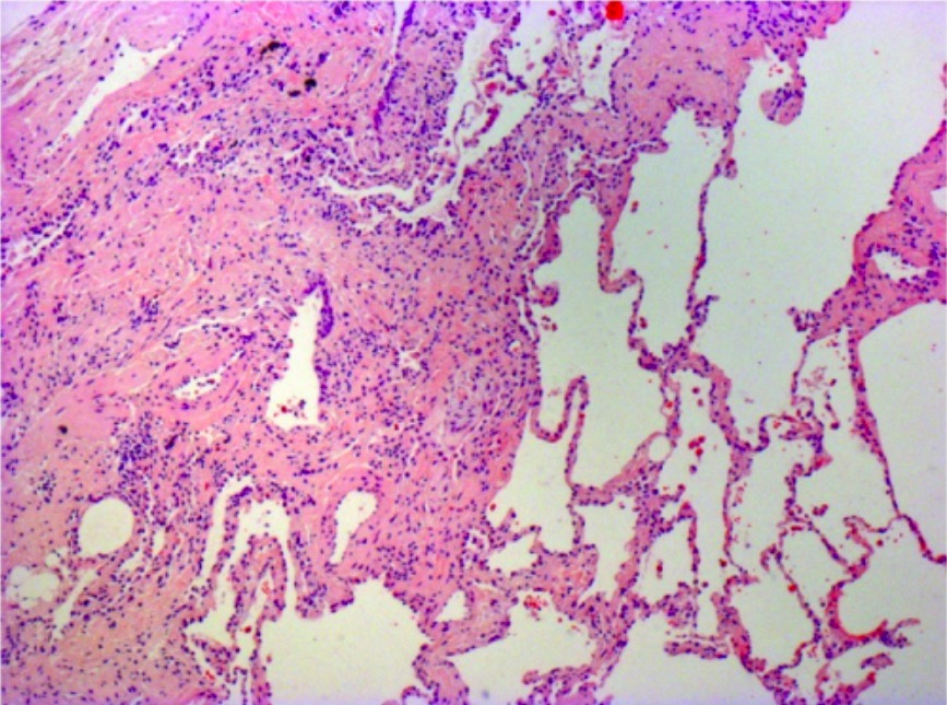
Photomicrograph of the affected lung lobe, showing the severe ... Download scientific diagram | Photomicrograph of the affected lung lobe, showing the severe emphysema. Smooth muscle hyperplasia is present in some alveolar walls, and around the bronchiole (upper ...
Label the photomicrogram of the trachea. - Brainly.com Label the photomicrogram of the trachea. Get the answers you need, now! mooretami1829 mooretami1829 07/20/2022 SAT High School answered • expert verified Label the photomicrogram of the trachea. 1 See answer Advertisement













Post a Comment for "43 label the photomicrogram of the lung"